Latvia, 2002
Ministry of Education and Science
University of Latvia
Association of Biology Teachers
phone: +371-7034860, +371-7334125, Fax: +371-7223801, email: ulkoro@latnet.lv
|
13th International Biology Olympiad
Latvia, 2002 Ministry of Education and Science
|
| 4 Kronvalda boulv., Riga LV-1586, LATVIA
phone: +371-7034860, +371-7334125, Fax: +371-7223801, email: ulkoro@latnet.lv | |
Length of the practical test - 60 minutes; 40 points
In the laboratory you must solve 3 tasks
Task 1 – Plant Systematics
Task 2 – Plant Anatomy
Task 3 – Plant Physiology
Task 1 – Plant Systematics
Materials and instruments
You will use the instrument set that you received upon registering for the 13thIBO!
You will also use other instruments and materials: samples No. 1-8, magnifying glass
You have 8 plants (samples 1-8) which can belong to the following plant families (A-J)
In the code table:
Code Table
|
A. Apiaceae |
E. Fabaceae | H. Poaceae |
|
B. Asteraceae |
F. Geraniaceae | I. Ranunculaceae |
|
C. Brassicaceae |
G. Lamiaceae | J. Rosaceae |
|
D. Araceae |
By morphological characteristics, determine the respective families (from those given in the code table), for the samples (1-8)!
Q1. Identify each sample in the dichotomous identification key below. Enter the sample number in the provided windows in the answer sheet. (8 points)
Write the family codes (A-J) in the windows provided beside the appropriate sample numbers!(8 points)
(continued on next page)
Identification key
| Thesis Nr. | Sample Nr. (1-8) | Family code
(A-J) |
|
| 1. | Flowers without perianth. Venation parallel … 2. | ||
| – | Flowers with calyx and corolla. Venation netted … 3. | ||
| 2. | Inflorescence spike | ||
| – | Inflorescence panicle as a dense cylinder | ||
| 3. | Inflorescence head | ||
| – | Flowers single or in an inflorescence, that is not a head … 4. | ||
| 4. | Flowers actinomorphic … 5. | ||
| – | Flowers zygomorphic … 7. | ||
| 5. | Leaves entire or lobed… 6. | ||
| – | Leaves separated. Inflorescence of compound umbels. G(2) | ||
| 6. | Ca5Co5A5+5G(5) | ||
| – | Ca2+2Co2+2A2+4 G(2) | ||
| 7. | Leaves opposite, entire; fruit – nutlet | ||
| – | Leaves alternate, compound; fruit – legume |
Task 2 – Plant Anatomy
Materials and instruments:
You will use the instrument set that you received upon registering for the 13thIBO!
You will also use other instruments and materials: sample No. 9, pith of black elderberry (for fixing in task 2), microscope, stain mixture, Petri Dish with water, distilled water, microscope, slides and coverslips, razor blade, filter paper, cloth material.
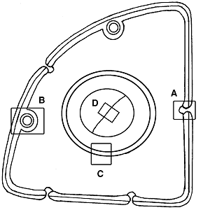
Cut the pith of black elderberry(Sambucus nigra) lengthwise in half with a razor blade. Holding with hands, secure sample No. 9 lengthwize between the halves (Figure 1. A-D). The black elderberry pith is used only for fixing. Holding the black elderberry pith in one hand and the razor blade in the other, prepare cross-sections of sample No. 9 (Figure1E) and place them in the water in the Petri Dish! Choose the three best cross-sections (without the black elderberry pith) and place them on the microscope slide. Add a drop of Astra blue (stains cellulose) and safranin (stains lignin) mixture. After 0.5 minutes, remove the stain with the filterpaper, and add a drop of distilled water, and remove them with the filter paper. Twice repeat rinsing with water. Add a drop of water and place the coverslip over the cross-sections.
The quality of the cross-section and the preparation will be assessed!
 |
Figure 1. Preparation of cross-section of sample No. 9 A – Black elderberry pith B – Cut black elderberry pith C – Sample placed between cut black elderberry pith D – Ready for cutting E – Preparation of cross-section |
Q 2 A. When the preparation is ready, using the low power x10 objective, find the best cross-section. Raise the card showing “2A”, and the assistant of the laboratory task will assess the quality of the cross-section and preparation, and write the assessment points in the answer sheet! (2 points)
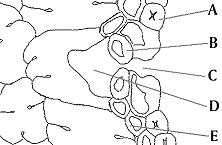
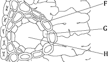
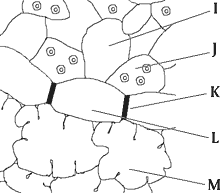
Q 2 B. Using the microscope (objective x10 and x40), study the preparations. Compare the cross-section seen under the microscope with that in Figure 2. and with its fragments [A (Figure 3), B (Figure 4), C (Figure 5), D (Figure 6)].
|
|
Figure 2. Cross-section of sample No. 9. | |
A. |
Figure 3. Sample No. 9 cross-section fragment A. |
|
B. |
Figure 4. Sample No. 9 cross-section fragment B |
|
C. |
Figure 5. Sample No. 9 cross-section fragment C | |
D. |
Figure 6. Sample No. 9 cross-section fragment D | |
Code Table
| No. | Part | No. | Part |
| 1. | Pericycle | 13. | Phloem |
| 2. | Cystolith | 14. | Transfusion tracheid |
| 3. | Transfusion parenhima | 15. | Palisade parenchyma |
| 4. | Hypodermis | 16. | Spongy parenhyma |
| 5. | Casparian strip | 17. | Guard cell |
| 6. | Endodermis | 18. | Sclerenchyma sheath |
| 7. | Back cavity | 19. | Raphide |
| 8. | Trichome | 20. | Angular collenchyma |
| 9. | Epidermis | 21. | Front cavity |
| 10. | Druse | 22. | Lobed parenchyma |
| 11. | Resin duct | 23. | Xylem |
| 12. | Epithelial cells | 24. | Pith |
In the answer sheet beside the letters A-O of parts seen on Figures 3-6, write the codes of the correct names of these parts!(15 points)
Q 2 C. Choose the correct plant taxon observed, and enter an “x” beside the respective code in the answer sheet.
Code table:
| A. | Bryophyta |
| B. | Equisetophyta |
| C. | Pinophyta |
| D. | Magnoliophyta |
(1 point)
Q 2 D. Choose the correct ecological group of the plant observed, and enter an “x” beside the respective code in the answer sheet.
Code table:
| A. | hydrophyte |
| B. | hygrophyte |
| C. | mesophyte |
| D. | xerophyte |
(1 point)
Task 3. Plant Physiology
Materials and instruments:
You will use the instrument set that you received upon registering for the 13thIBO!
You will also use other instruments and materials: sample No.10 – onion fragment, microscope, Ca(NO3)2solution, distilled water, microscope slides and coverslips, razor blade, filter paper, cloth material.
A characteristic stem modification (bulb) fragment from a representative of the Liliaceae is supplied. Separate from the bulb fragment one fleshy inner scale leaf, using the instrument set!
Q 3 A. Determine on which side of the fleshy inner scale leaf can be found the lower epidermis. Lift the card with sign “3A”, the assistant of the laboratory task will arrive, and you will show him the lower epidermis. His assessment will be entered in the answer page!(1 point)
Q 3 B. Make a preparation: using a razor blade, shave a thin (~5 x 5 mm) section of the lower epidermis and place it on the microscope slide. Add one drop of the Ca(NO3)2 solution, place a coverslip over the section, and begin observation of the process occurring immediately under the x10 objective. Raise the card “3B”, and the assistant of the laboratory task will arrive. His assessment of the preparation quality will be entered in the answer page! (1 point)
Q 3 C. What is the name of the process seen under the microscope? In the answer sheet, enter an “x” beside the code of the correct process.
Code table:
| A. |
Hemolysis |
| B. |
Dissociation |
| C. |
Association |
| D. |
Plasmolysis |
| E. |
Deplasmolysis |
| F. |
Hemophosphorylation |
(1 point)
Q 3 D. Which of the below concentrations of Ca(NO3)2 solution could have caused the process observed? Enter an “x” beside the correct codes of the possible concentrations in the answer sheet.
Code table:
| A. |
1 M |
| B. |
0.5 M |
| C. |
0.1 M |
| D. |
0.05 M |
| E. |
0.01 M |
(2 points)
| |||||
| |||||
| |||||
| Registration | |||||
| |||||
| Supports | |||||
| Results | |||||
|
Final Report
English version of tasks Russian version of tasks | |||||
| |||||
| Feedback | |||||
|
Monday October 18, 2004; 20:59
Main Frame | |||||